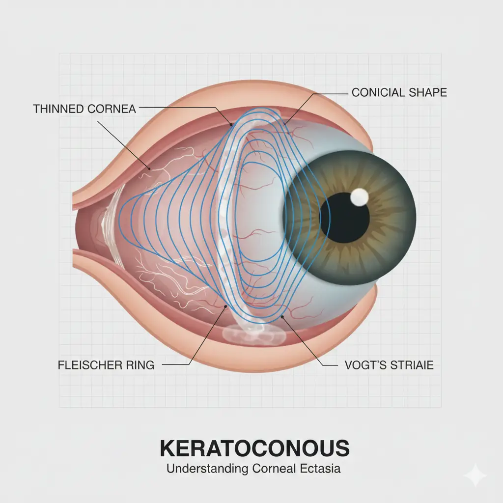
Keratoconus is a progressive eye condition in which the normally round cornea becomes thin and gradually bulges outward into a cone-like shape. This change distorts vision and can make everyday activities—like reading, driving, or using screens—difficult without proper treatment. Early diagnosis plays a major role in preventing long-term vision loss.
What Is Keratoconus?
The cornea is the eye’s clear, dome-shaped front surface that helps focus light.
In keratoconus, the corneal structure weakens, causing:
- Thinning of the cornea
- Bulging into a cone shape
- Irregular astigmatism
- Increasing blurry or distorted vision
The condition often begins during the teenage years and may progress until the mid-30s.
Common Symptoms of Keratoconus
Early symptoms may be mild, but they worsen as the cornea becomes more irregular. Patients may experience:
- Blurred or distorted vision
- Increased sensitivity to light and glare
- Difficulty seeing at night
- Frequent changes in glasses prescription
- Ghost images or double vision in one eye
- Eye irritation or headaches
If you notice constant prescription changes, it may be a warning sign of early keratoconus.
Causes & Risk Factors
Although the exact cause is unknown, several factors can increase the risk:
- Genetics: Family history of keratoconus
- Eye Rubbing: A major trigger that accelerates corneal thinning
- Allergies: Itchy eyes that lead to rubbing
- Connective Tissue Disorders: Such as Down syndrome or Marfan syndrome
Avoiding eye rubbing and managing allergies can help slow the progression.
How Is Keratoconus Diagnosed?
Eye doctors use advanced diagnostic tests, such as:
- Corneal Topography: Maps the shape and curvature of the cornea
- Pachymetry: Measures corneal thickness
- Pentacam / Orbscan: 3D imaging that detects early keratoconus
- Refraction Test: Checks visual clarity and prescription changes
These tests can detect keratoconus even in early stages, before symptoms become severe.
Treatment Options for Keratoconus
The right treatment depends on the stage and severity of the disease.
1. Glasses or Soft Contact Lenses (Early Stage)
Mild keratoconus can be managed with updated glasses or soft toric contact lenses.
2. Rigid Gas-Permeable (RGP) or Scleral Lenses
These special lenses create a smooth optical surface and provide clearer vision in moderate to advanced keratoconus.
3. Corneal Cross-Linking (CXL)
A breakthrough treatment that strengthens the cornea and stops the progression of keratoconus.
CXL involves:
- Vitamin B2 (riboflavin) drops
- UV light activation
- Formation of new collagen bonds to stiffen the cornea
It is currently the most effective treatment to prevent further damage.
4. Intracorneal Ring Segments (ICRS)
Small curved implants placed inside the cornea to flatten its shape and improve vision.
Useful in moderate cases where contact lenses no longer work well.
5. Corneal Transplant (Advanced Stage)
If the cornea becomes extremely thin or scarred, a partial or full corneal transplant may be required.
Modern techniques allow faster recovery and high success rates.
Lifestyle Tips to Protect Your Eyes
- Avoid rubbing your eyes
- Treat allergies promptly
- Use prescribed eye drops regularly
- Attend follow-up visits to monitor progression
Consistent care makes a huge difference in long-term outcomes.
When Should You See an Eye Specialist?
See an eye doctor if you experience:
- Frequent changes in glasses
- Blurry or distorted vision
- Increased glare or halos
- History of keratoconus in your family
Early intervention can stop progression and preserve vision.
Conclusion
Keratoconus is a manageable condition when diagnosed early. With modern treatments like Corneal Cross-Linking, scleral lenses, and ICRS, patients can maintain excellent vision and prevent the disease from worsening. If you or someone you know shows symptoms, consult an eye specialist for a proper evaluation.
Need Expert Medical Advice?
Our team of experienced doctors is here to help you with any health concerns.
Book Appointment